Diego Abreu, MS1, Shany Quevedo, MD2, Adalberto Gonzalez, MD3 1Universidad Iberoamericana, Santo Domingo, Distrito Nacional, Dominican Republic; 2Aventura Hospital Internal Medicine Residency, Aventura, FL; 3Cleveland Clinic Florida, Weston, FL
Introduction: Collagenous colitis is caused by an abnormally thickened subepithelial collagen deposition and inflammation in the colon. This leads to non-bloody diarrhea with nonspecific laboratory findings in middle aged patients. There may also be associated deposition of subepithelial collagen in the stomach (collagenous gastritis) or the small bowel (collagenous enteropathy). Protein losing enteropathy and iron deficiency anemia (IDA) have been infrequently associated with these conditions. We present a 56-year-old female with diarrhea for 1 month, protein losing enteropathy, and IDA.
Case Description/Methods: A 56-year-old healthy female presented for seven daily episodes of non-bloody, watery diarrhea for 1 month. She denied rectal bleeding, weight loss, or fever. Vital signs were normal. Physical exam showed mild dependent edema in bilateral ankles. Initial evaluation was with laboratory testing: WBC 4.3, Hb 7.7, MCV 76, Platelets 350, normal electrolytes, liver enzymes, creatinine, low albumin 2.9, low total protein 4.6. Normal celiac testing. Stool infectious testing was normal. Due to low protein, further testing was performed: Alpha-1-antitrypsin 635 mg/dL (low), Albumin 2.9 g/dL, total protein 4.6 g/dL, ferritin 17. Subsequently, esophagogastroduodenoscopy (EGD) and colonoscopy were recommended.
EGD revealed mucosal atrophy in the antrum, a normal esophagus and duodenum. Colonoscopy revealed mild loss of vascularity from the descending colon to the cecum. Pathology revealed markedly thickened collagen plate accompanying heavy lymphoplasmacytic infiltrate in the lamina propria from the cecum to the sigmoid colon; the rectum was not affected. There was also a thick collagen subepithelial deposition in the gastric antrum. Duodenal biopsies were normal. Diagnoses of collagenous colitis and collagenous gastritis were established. We prescribed oral Budesonide 9 mg daily for 2 months, then 6 mg for 2 weeks, then 3 mg for 2 weeks. Her diarrhea resolved within one week of starting budesonide. Laboratory studies had returned to normal after budesonide treatment.
Discussion: Our patient had an atypical presentation of collagenous gastritis and colitis due to a rapid onset of symptoms, protein losing enteropathy, and IDA. It was likely the involvement of the stomach leading to hypoacidity that led to the iron deficiency anemia and the protein losing enteropathy. Although we did not find evidence of collagenous enteropathy, it may cause similar laboratory findings due to malabsorption.
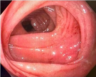
Figure: Colonoscopy images showing mild loss of vascularity in the mucosa of the colon.
Disclosures:
Diego Abreu indicated no relevant financial relationships.
Shany Quevedo indicated no relevant financial relationships.
Adalberto Gonzalez indicated no relevant financial relationships.
Diego Abreu, MS1, Shany Quevedo, MD2, Adalberto Gonzalez, MD3. P1700 - Collagenous Gastritis and Collagenous Colitis Presenting With Protein-Losing Enteropathy and Iron Deficiency Anemia, ACG 2023 Annual Scientific Meeting Abstracts. Vancouver, BC, Canada: American College of Gastroenterology.